Drilling down on the necessity of dental X-rays

- Share via
When my son and daughter were youngsters, once a year I’d have a disagreement with their pediatric dentist. He wanted to do routine annual X-rays, and I would protest because neither child ever had any cavities. His response: Dental X-rays are an important diagnostic tool, representing a small speck in the sea of radiation that we receive by inhabiting planet Earth.
It turns out we both were right. Dental X-rays are essential for detecting serious oral and systemic health problems, and generally the amount of radiation is very low. But new thinking on dental X-rays is that the “one size fits all” schedule is outdated.
“The notion of bite-wing X-rays every year and a full set of X-rays every three years for every patient should go in the garbage can,” says Stuart White, a dentist and professor emeritus at the UCLA School of Dentistry. Instead, decisions should be made individually.
Emphasizing that “without dental X-rays we would go back 120 years, and disease detection would be primitive and awful,” White says dentists must strive to minimize unnecessary exposure.
And this is where the discussion gets complicated because the amount of radiation you receive depends on how the dentist takes pictures of your teeth.
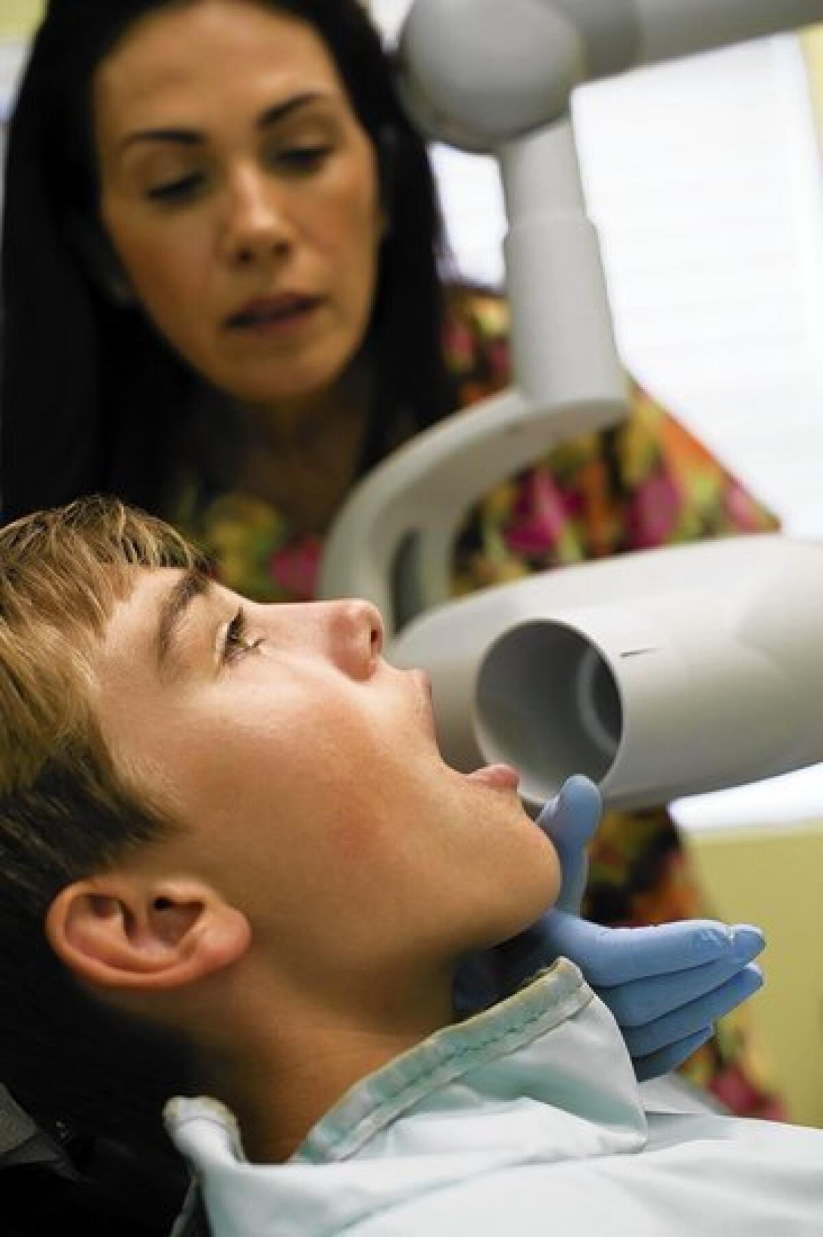
For example, if your dentist uses slow film and round collimation (the piece of equipment placed near your face during X-rays), you’re going to get approximately double the dose that you would from digital imagery and rectangular collimation.
“All dentists should follow the ALARA principle (as low as reasonably achievable) when determining what X-rays to order. Adhering to this principle will minimize the patient’s radiation exposure,” says Dr. Mark Kirkland, an associate dean at the UC San Francisco School of Dentistry.
So why would any dentist favor equipment that ups the dose of radiation?
“They’re used to one system and they don’t want to change it,” says Dr. Elham Radan, director of the radiology clinic at USC’s dentistry school.
But even dentists who haven’t switched to digital imagery can reduce patients’ risk by opting for faster film, Radan says, adding that “another important factor is that the technician who is taking the images is well-trained and skilled, which reduces the amount of re-exposures.”
As for round collimation, the problem is that it exposes more tissue, including the salivary glands, which are sensitive to radiation. Changing to rectangular collimation is not a big expense — systems can be converted for a few hundred dollars.
The ionizing radiation in X-rays is especially worrisome for children because their developing tissue is more vulnerable. There is concern about a new trend in orthodontics as popularity grows for cone-beam CT scanners, used to create 3-D images. Radiation doses from these machines can vary dramatically, depending on the manufacturer and settings.
Cone-beams are useful for oral surgeons, White says, but he criticizes routinely doing 3-D imaging when kids need braces.
Whenever any kind of dental X-ray is done, patients should wear protective lead aprons with thyroid collars. (However, the collar cannot be worn with all radiography, because sometimes it blocks the image.)
The aprons do not shield us from internal radiation “scattering,” especially with sensitive tissue like the thyroid gland. That’s why it’s important to reduce the X-ray beam to the smallest size that provides necessary information, says Sharon Brooks, a dentist and an expert at the nonprofit Health Physics Society, devoted to radiation safety.
Dental X-rays made news in 2012 after the American Cancer Society journal Cancer published a widely criticized study saying that people with one type of brain tumor remembered having twice as many dental X-rays as individuals in the control group, who did not have brain tumors.
Such studies, which rely on participants’ memories, can be problematic, says Dr. Otis Brawley, the society’s chief medical officer. He adds that, ideally, the controversial research will be used “to justify doing a larger, more definitive study, actually trying to show causality.”
But Brawley says the message of the study is valid: Dental X-rays should be done only when medically indicated, tailored to each patient’s health needs.


